What are the benefits of 4D Echocardiography?
- It’s An Advanced Ultrasound Test
- It Helps Resolve Heart Related Diseases
- It Can Identify Changes In The Heart
- It’s Developed From 3 Imaging Methods
- It Takes About An Hour To Complete
Perpetual Help Medical Center-Las Piñas, in partnership with our Heart and Vascular Institute, proudly presents the “Love Your Heart” event. PHMC is the only hospital in Las Piñas equipped with a 3D Echocardiography machine to better serve you as a patient. This machine can be beneficial to improve heart health and to properly diagnose patients with heart-related diseases. Before taking advantage of this opportunity, it’s important to learn about the benefits of 4D Echocardiography. Here are the following benefits you need to know.
It’s An Advanced Ultrasound Test
An echocardiogram is an advanced ultrasound test that creates images of the heart. Instead of using 3D technology, the 4D system creates a live video effect. This can also be used for prenatal tests. Since it will give you real-time images and videos, it will provide your doctor with an accurate evaluation of your heart. It will be easier to see its function and structure with this innovative technology. Even the condition of the heart chamber and valves can be seen using this test.
It Helps Resolve Heart Related Diseases
The images acquired from this test can assist in treating various heart diseases. Detailed representation and imaging assist in facilitating the analysis of coronary artery diseases, rhythmic disorders, and more. The test will help your doctor see the size, shape, thickness, and movement of your heart. It can depict how your heart moves and how strong it pumps. Doctors can see if your valves are working correctly or if there are any leaks.
The 4D ultrasound system also lets your doctor know if your heart valves are too narrow and if there are any abnormal growths or tumors around it. The following can also be found using 4D echocardiography:
- Problems with pericardium or outer heart lining
- Large blood vessels that enter and exit the heart
- Blood clots in heart chambers
- Holes between different heart chambers
It Can Identify Changes In The Heart

4D echocardiography is an excellent screening method. For example, patients who are using cardio-toxic medications for cancer treatment can get proper screening and diagnosis to see the health of the heart. If the patient has received radiation in the chest area, a 4D echocardiography can help see if the area is in normal condition. For patients with certain heart diseases such as cardiomyopathy, pericardial disease, and heart failure, this can be used to monitor their conditions. This is also especially helpful for patients with pacemaker implants.
It’s Developed From 3 Major Imaging Methods
There are 3 major imaging methods used to monitor the heart and strain rate. The first commercially available imaging method is the tissue image Doppler imaging. This method estimates velocity and strain in certain directions of the ultrasound. However, this method isn’t as effective as there are limitations. Out-of-plane motions are usually ignored by this method.
The next imaging method uses speckle tracking. This is an alternative approach to Tissue Doppler. While other methods remain static, the imaging of this uses motion analysis to see how different structures move from frame to frame. It’s easier to diagnose the image when speckles move together with the underlying structures of the heart. Speckle tracking is closer to the traditional methods of ultrasound recordings. Gray-scale recordings with the speckles are used to diagnose the condition of the heart accurately. However, out of plane motions are also a problem for this imaging method.
The last imaging method uses 3D block matching to assess 2D grayscale images while delivering motion estimates. The 4D strain is much more accurate while combining the same principles as the aforementioned imaging methods.
It Takes About An Hour To Complete

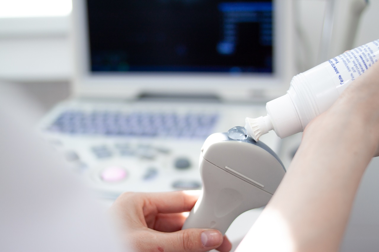
To get your echocardiogram, your doctor or sonographer will help you prepare. Remove clothing from the waist up and change into a hospital gown. You will lie on a table and a technician will proceed to add electrodes on your chest. These disks contain wires that communicate with the electrocardiograph machine. This will keep track of your heartbeat during the test.
The lights will be off during the test so the technician can see the video clearly. First, a gel will be applied to the chest to help sound waves pass through the skin easily. The technician may ask you to move or hold your breath to help get better images. The transducer will pass across your chest to probe the sound waves. These sound waves will then bounce off your heart and echo back to the transducer. This is how the image is created. Depending on the sound waves, the image will be shown as a photo or video on the monitor. The images created will be recorded for analysis later.
Key Takeaway
The benefits of 4D echocardiography can help a lot of patients who are suffering from heart-related diseases. It’s a one hour process that’s perfectly painless and can be done without much adjustment to your schedule. Visit PHMC’s Heart and Vascular Institute to get your own analysis. At PHMC, we strive to provide the best services to our patients.